Neonatal Neurology: Pre-Clinical Research
Neonatal Neurology 8: Preclinical 2
26 - Microglial activation after IUGR – is it friend or foe?
Publication Number: 26.434
- CF
Camille Fung, MD (she/her/hers)
Associate Professor
University of Utah
Salt Lake City, Utah, United States
Presenting Author(s)
Background:
Offspring both with intrauterine growth restriction (IUGR) are at increased risk for learning and memory deficits. Abnormal hippocampal dendrite development seen as reduced arborization and synaptic formation is one proposed mechanism for memory deficits. Microglia are known to modulate dendritic/synaptic remodeling in the developing and adult brain, yet their role in IUGR is not well understood.
Objective:
To replicate reduced dendritic arborization presented in last year’s PAS but additionally determine the abundance and localization of microglia in the IUGR hippocampal dentate gyrus (HDG), the first relay station into the hippocampus proper for memory consolidation.
Design/Methods:
IUGR mouse pups were born after a potent vasoconstrictor, U-46619, infusion to mimic hypertensive disease of pregnancy in the last week of C57Bl/6J dams. Sham-operated dams gave birth to normally-grown pups. On postnatal day (P) 21, we labeled sham and IUGR neurons with a recombinant adeno-associated virus conjugated to green fluorescent protein (GFP) driven by the CAMKII promoter. At P28, we harvested brains and performed double immunofluorescent labeling for GFP+ dendrites and Alexa-594-labeled ionized calcium-binding adapter molecule 1+ (Iba1+) microglia. We enumerated dendritic branching and volumes, principal dendrite lengths, % Iba1+ immunofluorescence (IF) per unit area, Iba1+ cell sizes, and co-localization of Iba1+ cells with dendrites. Non-parametric Kruskal Wallis test was used to compare 4 groups (n=3-4/group).
Results:
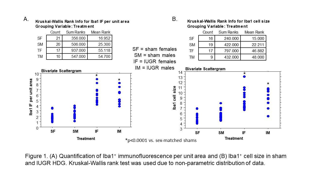
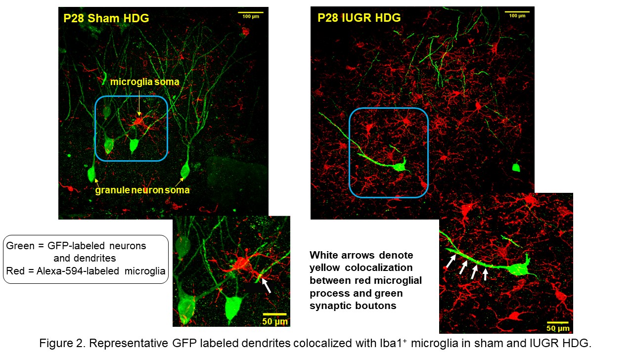
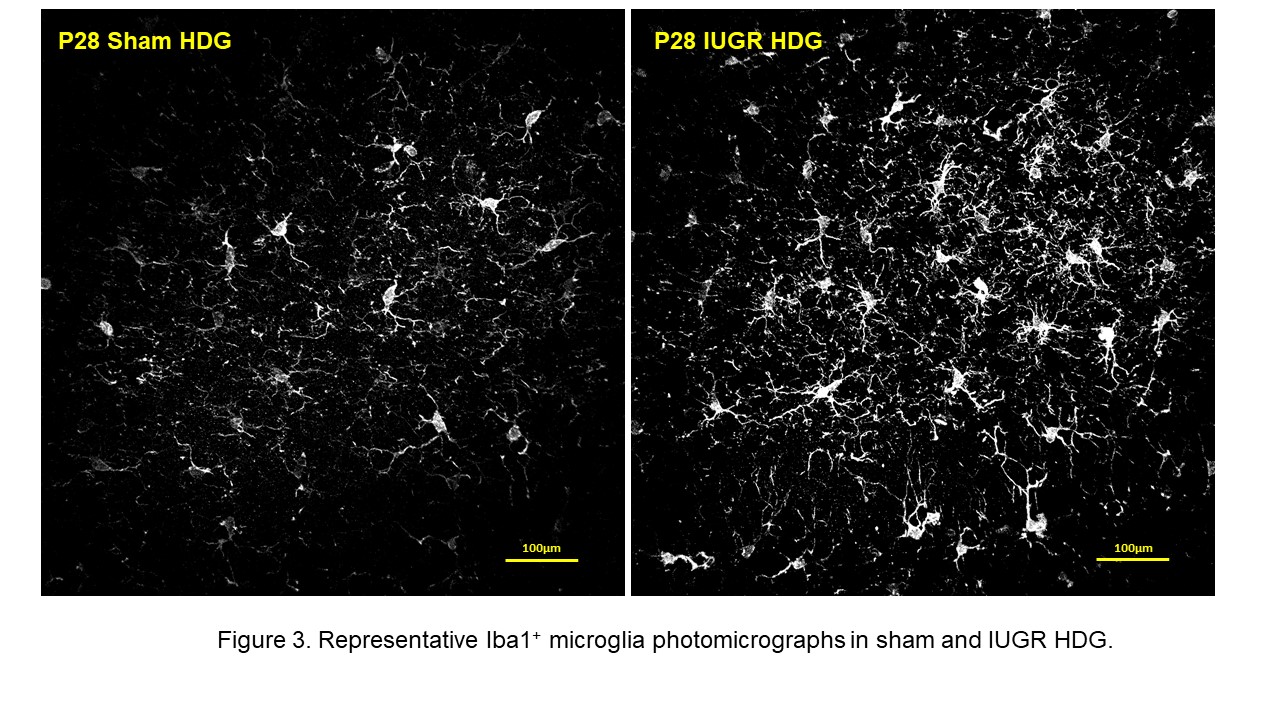
Similar to our findings from last year, we replicated decreased dendritic branching and volumes in the IUGR male and female HDG and found similar principal dendrite lengths compared to sex-matched shams (data not shown). More importantly, we found a 3.2x and 2.4x increase in Iba1+ IF/unit area in IUGR female and male HDG respectively compared to their shams (p< 0.0001, Figure 1A). Iba1+ cell bodies were 1.6-1.7x larger in IUGR males and females with shorter and thicker ramifications (p< 0.001, Figures 1A, 2, 3). Activated Iba1+ microglia were localized to synaptic boutons in IUGR HDG (Figure 2).
Conclusion(s):
IUGR increased HDG microglial number and altered cell morphology consistent with microglial activation. This could explain the reduced dendritic arborization in IUGR. Interestingly, microglial activation can induce either a pro-inflammatory neurotoxic or anti-inflammatory neuro-reparative response. We plan to characterize pro- and anti-inflammatory cytokines as well as chemokines to characterize the microglial milieu in IUGR. 


