Emergency Medicine: All Areas
Emergency Medicine 12
501 - The Utility of Common Bile Duct Measurement in the Diagnosis of Cholecystitis and Choledocholithiasis in Pediatric Patients
Publication Number: 501.311

Marci Fornari, MD (she/her/hers)
Pediatric Emergency Medicine Attending
Children's National Health System
Washington, District of Columbia, United States
Presenting Author(s)
Background:
The incidence of gallbladder pathology in children is increasing. One component of biliary ultrasound (US) is measuring the common bile duct (CBD) which is normally < 4mm in children; this can be technically challenging to identify on biliary ultrasound US. No prior studies explore the utility of identifying the CBD as an essential component of biliary US to identify pathology in children.
Objective: Our primary outcome was to determine the presence of isolated CBD dilation in pediatric patients diagnosed with cholecystitis or choledocholithiasis with normal laboratory values and an otherwise unremarkable biliary US.
Design/Methods:
We performed a retrospective review of patients ≤ 21-years-old, at a single free-standing tertiary care children’s hospital, who received a biliary US in the radiology department from September 2005 to February 2020. We identified patients who had a radiology ultrasound (RADUS) diagnosis of acute cholecystitis and/or choledocholithiasis. We included patients who had cholecystectomy with surgical pathology and/or an endoscopic retrograde cholangiopancreatography (ERCP), as this is the gold standard for diagnosis of cholecystitis and/or choledocholithiasis. We excluded patients who did not have the CBD mentioned in the radiology report, labs within 14 days, or had a prior cholecystectomy. Prior adult studies defined a “positive” US as having any of the following: gallbladder wall thickening (GWT), pericholecystic fluid (PCF), and/or sonographic Murphy’s sign (SMS). We used SPSS to analyze frequencies for categorical variables, and medians and interquartile ranges for continuous variables.
Results:
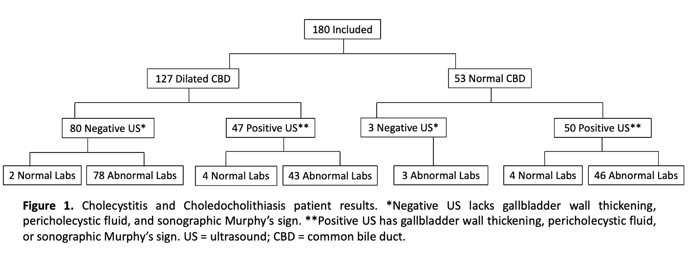
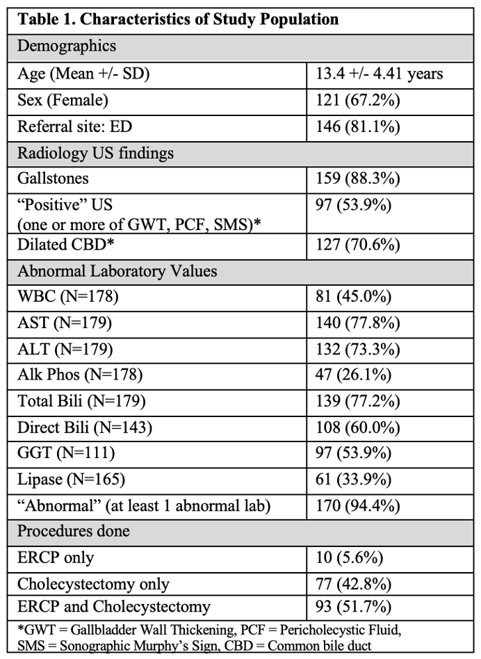
180 patients met inclusion criteria, with 121 (67.2%) female patients. Table 1 indicates the demographics of included patients. Table 2 indicates the cohort classification with percent that had abnormal labs and dilated CBD. Eighty one percent of the patients presented from the emergency department. Isolated CBD dilation without GWT, PCF or SMS or laboratory abnormalities occurred in 2 (1.1%) cases (Figure 1). Notably, those 2 patients did have an US diagnosis of choledocholithiasis.
Conclusion(s):
The prevalence of isolated sonographic CBD dilation in pediatric patients diagnosed with cholecystitis and choledocholithiasis was 1.1%. The two patients with isolated CBD findings with normal labs and “negative” ultrasound did have a RADUS diagnosis of choledocholithiasis. Thus, omission of CBD measurement is unlikely to result in missed diagnoses that require procedural intervention with an otherwise normal US and normal laboratory values. 


