Neonatal Neurology: Clinical Research
Neonatal Neurology 6: Clinical 6
130 - Corpus callosum abnormalities at term-equivalent age are associated with language development at two years corrected age in infants born very preterm
Sunday, April 30, 2023
3:30 PM - 6:00 PM ET
Poster Number: 130
Publication Number: 130.338
Publication Number: 130.338
Katsuaki Kojima, Cincinnati Children's Hospital Medical Center, Cincinnati, OH, United States; Julia Kline, National Institutes of Health, Bethesda, MD, United States; Mekibib Altaye, Cincinnati Children's Hospital Medical Center, cincinnati, OH, United States; Nehal A. Parikh, Cincinnati Children's Hospital Medical Center, Cincinnati, OH, United States

Katsuaki Kojima, MD, PhD, MPH (he/him/his)
Assistant Professor
Cincinnati Children's Hospital Medical Center
Cincinnati, Ohio, United States
Presenting Author(s)
Background: Very preterm infants (VPI) are at high risk for language impairment. Microstructural white matter abnormalities in the corpus callosum (CC) on diffusion tensor imaging (DTI) predict developmental impairment. It is unknown if this relationship is independent of clinical and social predictors.
Objective: To investigate the relationship between microstructural white matter abnormalities in the CC and language outcomes in VPI. We hypothesize that the fractional anisotropy (FA) of the anterior midbody of the CC predicts language development at age two, independent of clinical and social risk factors.
Design/Methods: We prospectively studied 348 infants born at or below 32 weeks' gestation from a geographically defined region. All participants completed DTI at term-equivalent age on a single 3T Philips Ingenia scanner and developmental assessments (Bayley-III) at two years corrected age. DTI was acquired using a 36-direction spin echo EPI sequence: TE 88 ms, TR 6972 ms, FOV 160 × 160 mm, in-plane resolution=2mm x 2mm; slice thickness=2mm; b-value of 800 s/mm2. Fraction anisotropy (FA) values of white matter tracts, including six regions of the CC, were calculated using TractSeg software. We developed multivariable linear regression models to evaluate the association between the mean FA values in the anterior midbody of the CC and Bayley language composite scores. We included predictors of language development, including sex, gestational age, postmenstrual age at DTI, high-risk social status based on the composite social risk score, severe brain injury based on the global brain abnormality score, late onset sepsis, and severe BPD (grade 3) as covariates in the multivariable models.
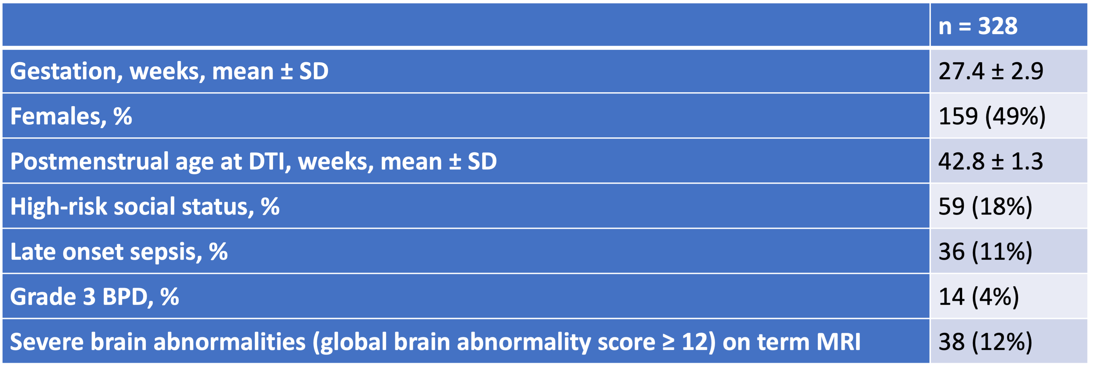
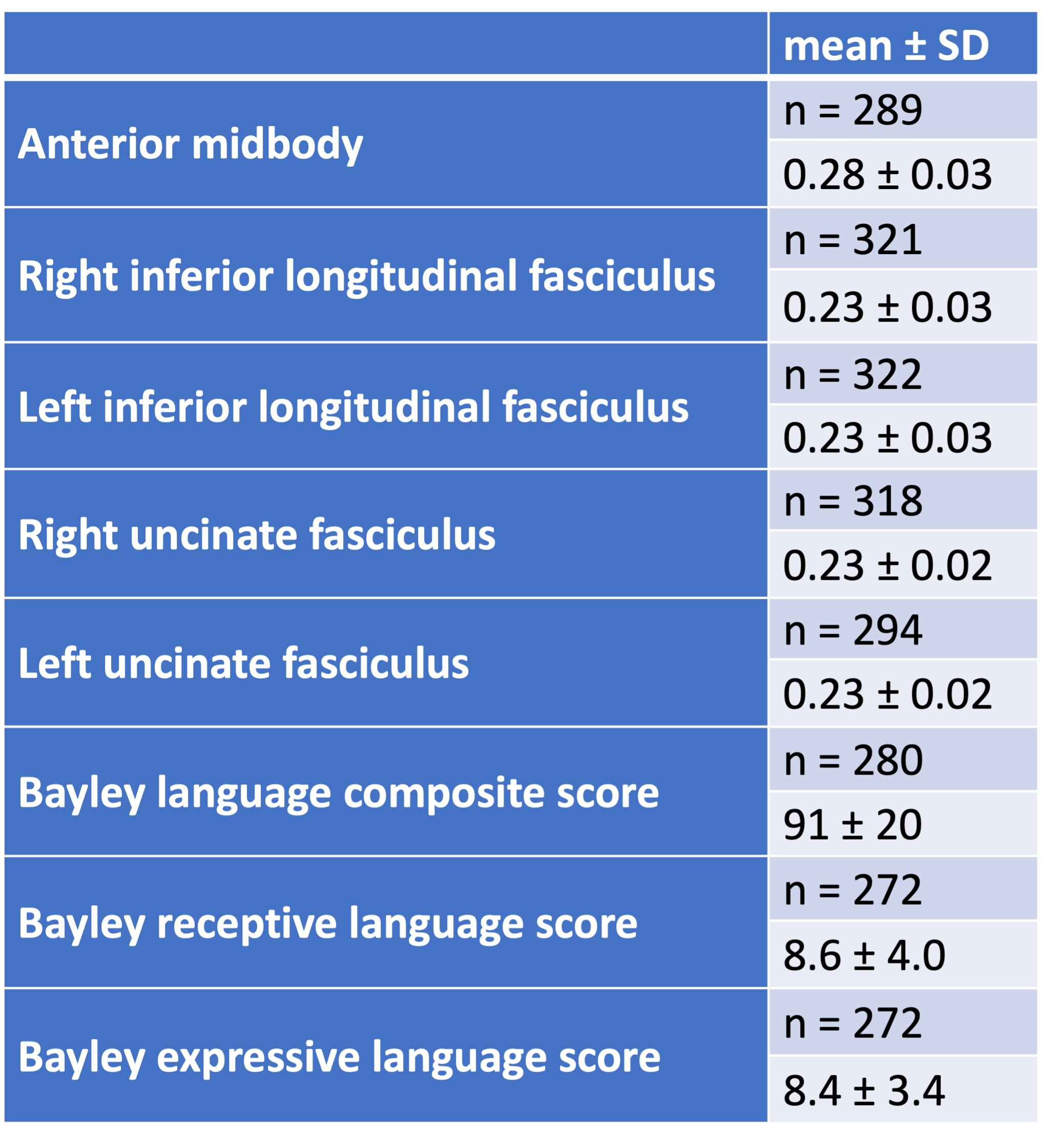
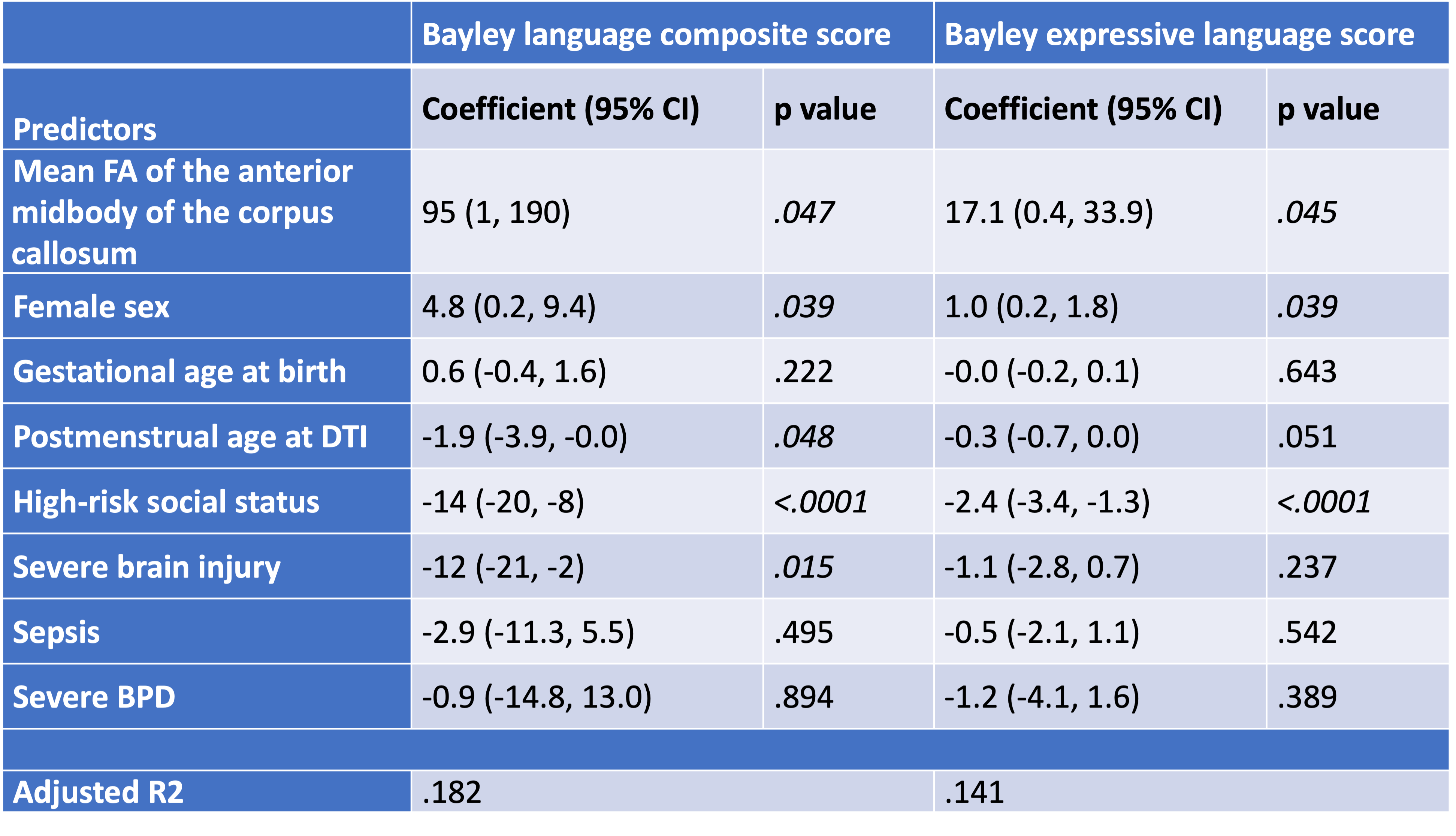
Results: Of the 348 infants in the study, 328 (94%) had high-quality DTI, and 280 (80%) completed the Bayley Scales assessment (Tables 1 and 2). Multivariable models showed that mean FA values in the anterior midbody of CC were significantly predictive of Bayley language composite scores (p = .047, adjusted R2: .182) and expressive language scores (p = .045, adjusted R2: .143) (Table 3). Mean FA values from the ILF and UF were not independent predictors of Bayley language scores at age 2.
Conclusion(s): In this regional cohort, FA of the anterior midbody of the CC was a significant predictor of language outcomes at two years corrected age in VPI independent of multiple clinical and social risk factors. Despite advances in detecting microstructural injury, predicting language development remains challenging.


