Neonatal Infectious Diseases/Immunology
Neonatal Infectious Diseases/Immunology 1
653 - Multi-Omic Analysis of Activation States in Circulating T cells in Very Preterm Infants
Publication Number: 653.24

Oluwabunmi Olaloye, M.D
Instructor of Pediatrics
Yale School of Medicine
Middletown, Connecticut, United States
Presenting Author(s)
Background:
Extremely premature infants (EPI) are susceptible to infections and inflammatory diseases. Helper(CD4) and cytotoxic(CD8) T cells are crucial for antigen-specific responses. Little is known about the ontogeny of circulating T cells in EPI.
Objective:
To define T cell ontogeny in EPI through multi-omic analysis of circulating immune cells in the first two months.
Design/Methods:
Serial blood samples of 0.25 ml each were obtained in the first 2 months of life from infants born at Yale New Haven Hospital (GA 25-30 weeks, n=10, Table 1). Cord blood samples from preterm (GA 29 – 35 weeks, n=5) and term infants (GA 37 – 39 weeks, n=7) and healthy adults (n=5) were included for comparison. Blood spots were analyzed with Olink ® proteomics. Circulating immune cells were analyzed by mass cytometry by time of flight (CyTOF) focusing on phospho-proteins and single-cell RNA sequencing (scRNAseq).
Results:
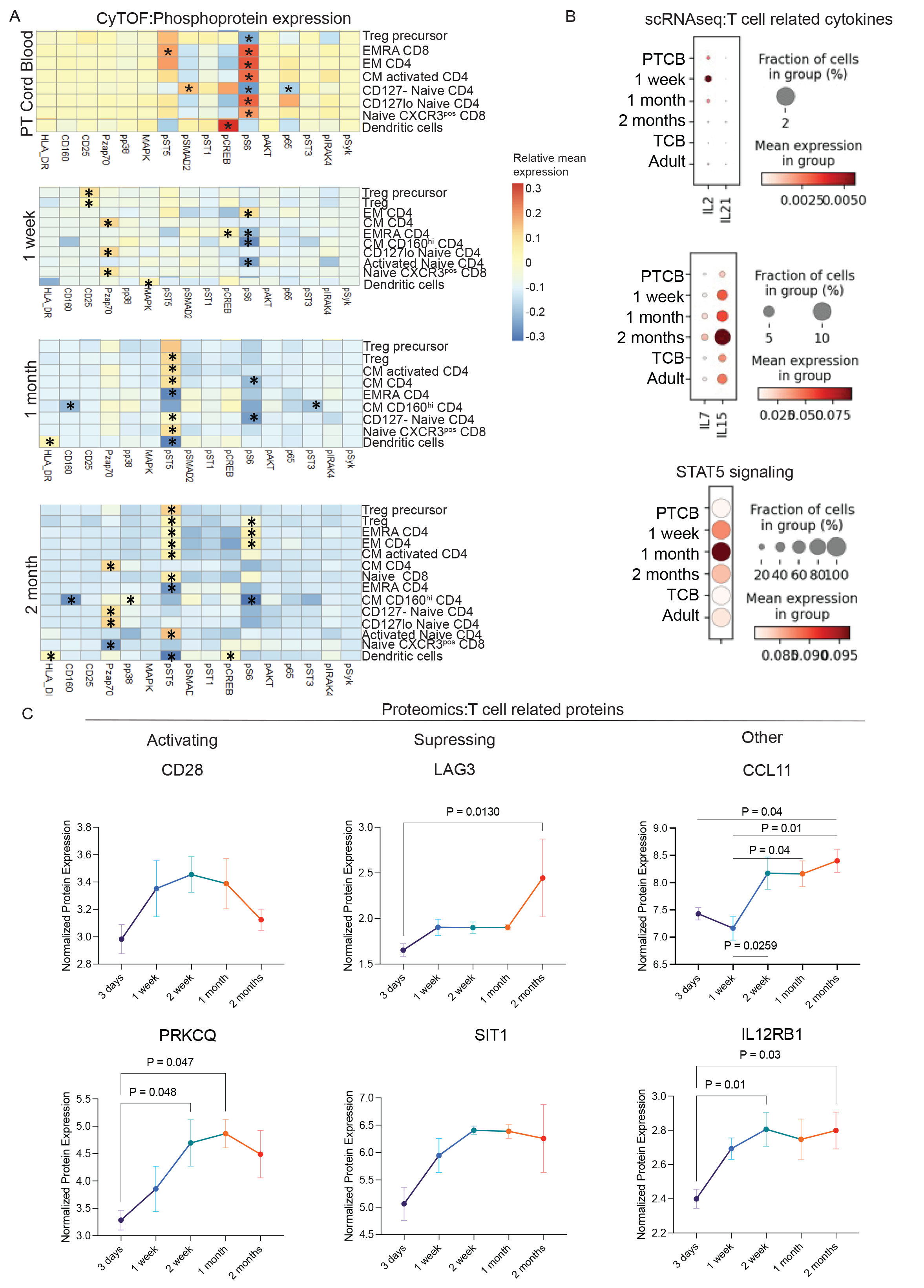
CyTOF and scRNAseq analysis identified several dynamic populations of circulating immune cells (Fig 1A-B) Differences in T cell abundance, phosphoprotein expression, transcriptome and clonality were noted. Naïve CD4 and regulatory T cells peaked at 1 month, while naïve CD8 T cells continuously increased. Central memory (CM) CD4 T cells were detected in cord blood and decreased from birth to 1 month. Meanwhile, effector memory (EM) CD4 and CD8 T cells were low at birth and increased by two months of age (Fig 1C-G). scRNAseq demonstrated an expansion of cycling T cells at 1 and 2 months of age (Fig 1H) and clonal expansion in memory CD8 T cells (Fig 1I). Protein phosphorylation levels in T cell populations were also dynamic (Fig 2A). PI3K/mTOR pathway (pS6) was suppressed at 1 month and increased at 2 months of age in memory T cell populations. TCR activation (pZAP70) was high in naïve and CM CD4 and CD8 populations concomitant with high dendritic cell activation as measured by HLA-DR, pCREB and pMAPK, suggesting ongoing antigen presentation and memory T cell generation (Fig 2A). pSTAT5 levels increased at 1 and 2 months of age consistent with more cycling T cells. scRNAseq data confirmed increased STAT5 signaling and transcription of cytokines essential for T cell activation and proliferation (IL2, IL7, IL15, Fig 2B). Finally, proteomic analysis showed changes in proteins involved in T cell activation, suppression, and function (Fig 2D).
Conclusion(s):
In EPI, there is a dynamic progression of T cell signaling, transcriptome, and clonality demonstrating activation, proliferation, antigen presentation, and memory formation. Thus, even EPI can mount antigen-specific adaptive immune responses early in life. 

