Neonatal Nephrology/AKI
Nephrology 6: Glomerular/Clinical and Basic Science
74 - Neonatal hyperoxia alters podocyte HIF-1α levels and induces podocyte senescence and cytoskeletal derangement
Sunday, April 30, 2023
3:30 PM - 6:00 PM ET
Poster Number: 74
Publication Number: 74.351
Publication Number: 74.351
Joanne Duara, University of Miami, Mia, FL, United States; Pingping Chen, University of Miami, Miami, FL, United States; Jeffrey D. Pressly, University of Miami, Miami beach, FL, United States; Haley Gye, University of Miami Leonard M. Miller School of Medicine, Potomac, MD, United States; Augusto F. Schmidt, University of Miami Miller School of Medicine, Miami, FL, United States; Shu Wu, University of Miami Leonard M. Miller School of Medicine, Miami, FL, United States; April W. Tan, University of Miami Leonard M. Miller School of Medicine, Miami, FL, United States; Merline Benny, University of Miami Leonard M. Miller School of Medicine, Miami, FL, United States; Alessia Fornoni, University of miami, Miami, FL, United States; Karen Young, University of Miami, Miami, FL, United States
- JD
Jo Duara, MD, MPH (she/her/hers)
Assistant Professor
University of Miami
Miami, Florida, United States
Presenting Author(s)
Background: Recent evidence suggests that podocyte loss may contribute to renal dysfunction in preterm infants. The mechanisms contributing to podocyte depletion are however unclear. Emerging data show that HIF-1α regulates podocyte cytoskeleton integrity, cell cycle, and function. Whether neonatal hyperoxia contributes to podocyte loss in preterm infants by altering podocyte HIF-1α levels, inducing podocyte senescence, and destabilizing the podocyte cytoskeleton is unknown.
Objective: To determine whether neonatal hyperoxia exposure decreases podocyte HIF-1α expression and induces podocyte senescence and cytoskeletal derangement as evidenced by decreased synaptopodin expression.
Design/Methods: In vitro, immortalized human podocytes were exposed to hyperoxia (85% oxygen) or normobaric normoxia for 24 hours. Cell proliferation was assessed by Ki67 stainin and ell senescence was evaluated by β-gal staining. In vivo, neonatal rats were randomly assigned to normoxia (RA) or hyperoxia (85% O2) from postnatal day 1 to 10. Following this exposure, urine protein/creatinine ratio was assessed, and kidney sections were immunostained with HIF-1α and synaptopodin antibodies.
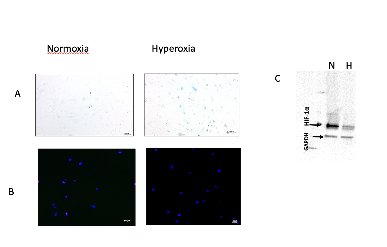
Results: In vitro, podocytes exposed to hyperoxia for 24 hours exhibited decreased cell proliferation (66% vs 33% per high power field) and increased senescence as evidenced by increased β-gal staining a[Fig 1 a, b]. In addition, Western blot analysis revealed that podocytes exposed to hyperoxia had 66% decrease in HIF-1α expression [Fig 1 c]. In vivo, pups exposed to 10 days of hyperoxia had increased urine protein/creatinine ratio (9 in the hyperoxia group vs 4.2 in the normoxia group) along with decreased HIF-1α and synaptopodin expression [Fig 2].
Conclusion(s): These findings suggest that neonatal hyperoxia alters HIF-1α levels, increases proteinuria, induces podocyte senescence, and deranges the podocyte cytoskeleton. Strategies targeting the HIF-1α pathway may have therapeutic benefits in attenuating neonatal kidney injury.

