Neonatal Infectious Diseases/Immunology
Neonatal Infectious Diseases/Immunology 3
404 - Immunohistochemical Evidence of Complement Activation in Stage III Necrotizing Enterocolitis Correlates with Preference for Endothelial Blood Group A Antigen Expression
Publication Number: 404.334

Kimberly N. Baran, DO (she/her/hers)
Neonatal Fellow
Loyola University Chicago Stritch School of Medicine
Wood Dale, Illinois, United States
Presenting Author(s)
Background:
Necrotizing enterocolitis (NEC) is a complex gastrointestinal disease affecting mostly preterm infants. The pathogenesis of this disease process is incompletely understood and thought to be multifactorial due to ischemia, infection, inflammation, endothelial injury, and immune dysfunction [1]. The complement system is a component of innate immunity for immediate recognition and elimination of invading microorganisms. It is non-specific, making no distinction between self and non-self, so its activation is very tightly controlled [2]. C4d is a split product of complement activation (CA) by either the classical (CP) or lectin pathways (LP), and binds tightly to the tissues at the site of activation. [2, 3]. Histologic changes to the bowel associated with NEC include coagulative necrosis, bacterial overgrowth, and cellular inflammation; however, immunohistochemical (IHC) properties of NEC have yet to be determined [1].
Objective: We sought to use C4d staining to identify if there is morphologic evidence of CA in surgical NEC tissues supporting possible antibody-mediated tissue injury.
Design/Methods:
Intestinal tissue samples were obtained from 12 infants (born 24-36 weeks gestational age) who underwent surgery for Bells Stage III NEC between 1.5-11.5 weeks of life. Viable, non-necrotic small bowel tissues were stained with rabbit monoclonal antihuman C4d antibody (Santa Cruz Biotechnology, Dallas, TX). Grading of C4d deposition is based on a scale widely accepted in transplant medicine [4]. Endothelial C4d staining was graded 0 (negative), 1 (minimal), 2 (focal), or 3 (diffuse). The Kruskal Wallis test compared C4d grade among the 4 different blood types. Post-hoc pairwise comparisons of C4d grade between each pairwise combination of blood types were performed.
Results:
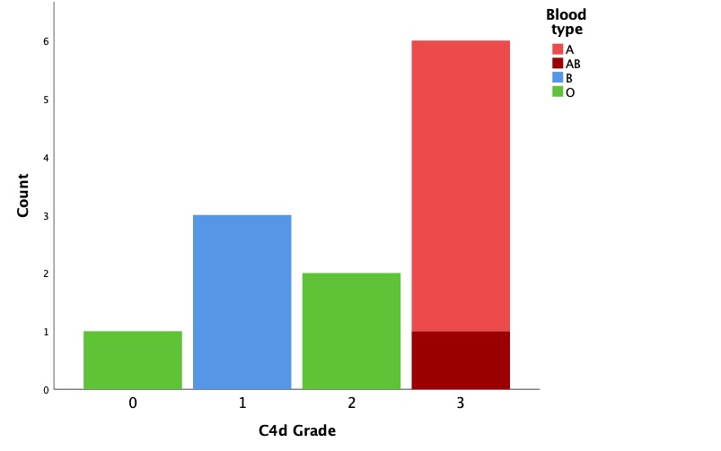
Figure 1 shows the frequency of C4d grade for each infant's tissue specimens within each blood type. All type A and type AB specimens had a grade of 3. All type B specimens had a grade of 1. Type O specimens had grades of 0 or 2. Median C4d grade differed between blood types (Table 1, p=0.02).
Conclusion(s):
Intestinal endothelia of infants with Bells Stage III NEC known to express blood group A antigen (whether blood type A or AB) bound C4d preferentially, suggesting increased CA in these infants as compared to infants with blood types B or O. The preference noted in this study may be related to the relative endothelial expression of A and B antigens. It is important to note that there was some staining of C4d in type O tissues, which is possibly due to CA by carbohydrate ligands of microorganisms via the LP.

