Hypertension
Hypertension 2
26 - Intrapulmonary Right to Left Shunt via Bronchial Circulation and Bronchial Endothelial Dysfunction in Experimental Pulmonary Hypertension Animal Models.
Publication Number: 26.322

Gregory Seedorf, BS (he/him/his)
Research Manager
University of Colorado School of Medicine
Aurora, Colorado, United States
Presenting Author(s)
Background:
Pulmonary hypertension (PH) is a devastating disease that effects many children and adults causing progressive right heart failure, premature death with no known cure. The bronchial circulation (BC) is vital in lung health as it provides oxygen and nutrition. However, its role in pulmonary vascular disease has been largely overlooked. Intrapulmonary bronchopulmonary anastomoses (IBA), which are vascular connections between the BC and PC are open in fetal life, closed after birth and when recruited it allows deoxygenated blood to bypass alveolar capillaries, leading to intrapulmonary right to left shunt (IRLS), hypoxemia and death. Importantly, the BC has been shown to contribute to plexiform lesions in PH, but whether IRLS utilizing the BC plays a role in PH has not been investigated.
Objective:
We hypothesize that in PH, there is increased pulmonary to bronchial flow that contributes to IRLS, which is partly mediated by abnormal bronchial endothelial cells (BEC) vasomotor function in neonatal and adult PH models.
Design/Methods:
Adult Sprague Dawley rats were exposed to weekly injections of SU5416 (100mg/kg) with continuous hypoxia (14% oxygen) for 5 weeks. At 5 weeks, green ink was injected into either the main pulmonary artery (MPA) or into the aorta (AO). Neonatal Sprague Dawley rats were exposed to a single dose of Su5416 at day of life 4 and ink or barium was injected into the MPA or AO at day 14, respectively. BEC were isolated from large airways by positive selection with CD31-coated magnetic bead and were studied at passage 1-2. Western blots were performed on BEC lysates for endothelin-1 (ET-1), adrenergic receptor (AR) and endothelial nitric oxide synthase (eNOS) function.
Results:
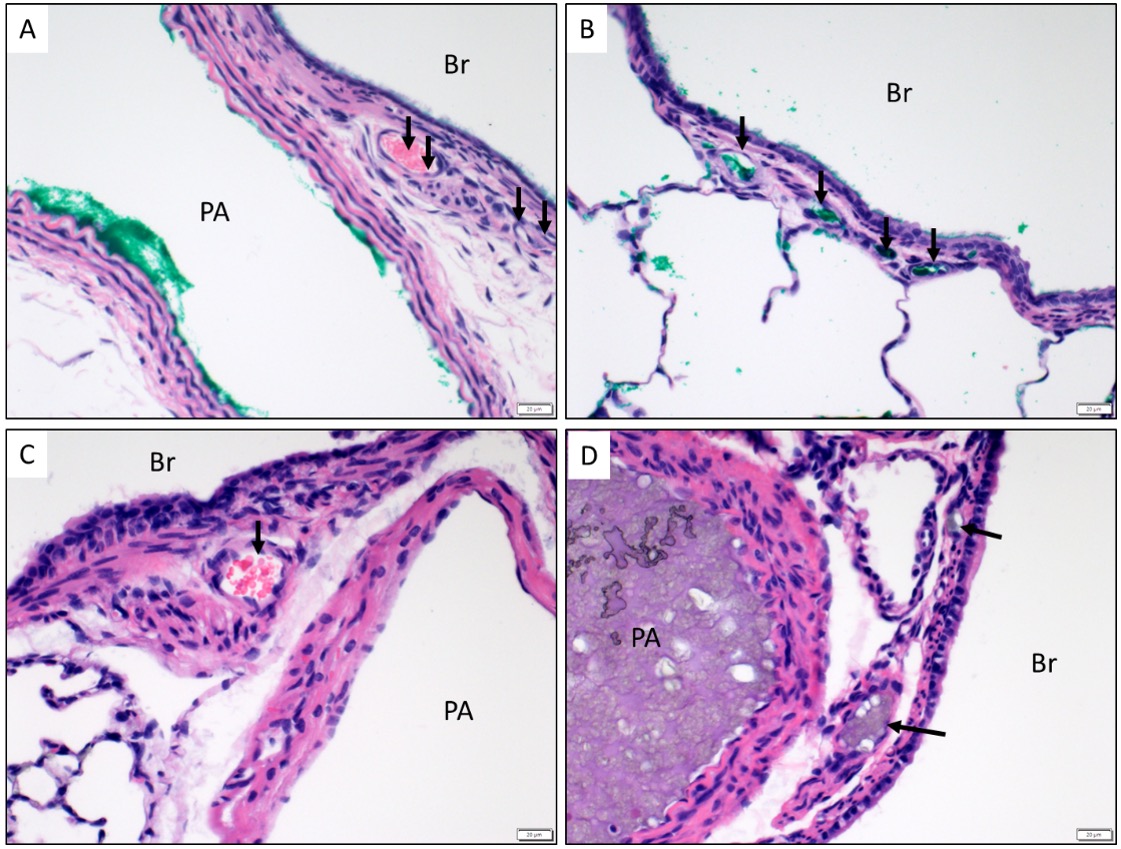
MPA injections of adult and neonatal PH rats show marked bronchial vessel filling and barium injection of MPA of neonatal PH rats also show significant bronchial vessel filling suggesting extensive IRLS via BC (Figure 1). Injections via AO do not identify green ink in PC, excluding left to right shunting. BEC isolated from SuH-treated rats show dysregulated function characterized by decreased ET-1 signaling, increased AR activity, and altered NO production.
Conclusion(s):
Neonatal and adult PH rats develop IRLS via PC to BC, and their BEC exhibit vasomotor dysfunction. These findings support past human data of IBA recruitment in neonatal and adult PH patients, suggesting that altered BC may contribute to the pathobiology of PH.
