Neonatal Neurology: Pre-Clinical Research
Neonatal Neurology 9: Preclinical 3
32 - Effect of preterm hyperbilirubinemia on adolescent rat cerebellum
Publication Number: 32.435
.jpg)
Cansu Tokat, MD (she/her/hers)
Research Assistant
Case Western Reserve University School of Medicine
Shaker Heights, Ohio, United States
Presenting Author(s)
Background:
Bilirubin is produced by the breakdown of hemoglobin and is normally catabolized and excreted. Accumulation of bilirubin can become neurotoxic, as is often the case in premature infants. The homozygous Gunn rat (jj) lacks uridine diphosphate glucuronosyltransferase 1A1 (UGT1A1), the enzyme needed to biotransform bilirubin. jj rats can be made acutely hyperbilirubinemic by injection of sulfadimethoxine (sulfa). This drug displaces bilirubin from albumin, and thus increases free bilirubin which can cross the blood brain barrier and cause neurotoxicity. Heterozygous littermates (Nj) have reduced UGT1A1 (~50% of wild type), do not develop hyperbilirubinemia and serve as controls.
Objective:
The objective of this study was to determine if changes in the cerebellum could be seen on Magnetic Resonance Imaging (MRI) of adult animals made hyperbilirubinemic on a postnatal day 5.
Design/Methods:
Gunn rat pups were randomly assigned to be injected intraperitoneally with either sulfa (200 mg/kg) or an equivalent volume of saline (sal) on a postnatal day 5 (P5). A 7T Bruker Biospec scanner with a 30cm bore and 400 mT/m magnetic field gradients and a 35mm ID mouse volume coil for signal detection were used to acquire T2-weighted images on postnatal day 37 (P37). P37 is considered to be the adolescent/young adult equivalent of human central nervous system. A region of interest analysis was performed at the level of the middle cerebellar peduncle (MCP) of the cerebellum and brainstem. The volumes of these areas were measured via Paravision 5.1 software.
Results:
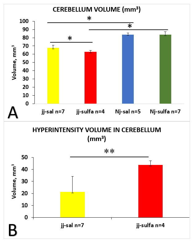
Cerebellar volume analysis showed: 1) No difference in volume between Nj-sal and Nj-sulfa; 2) A significant decrease in volume in jj-sal compared to Nj-sal; 3) A significant decrease in volume in jj-sulfa compared to jj-sal (Figure 1A). jj rats only exhibited white matter changes as significantly increased areas of hyperintensity. The volume of the hyperintense areas at the level of the MCP was significantly increased in jj-sulfa compared to jj-sal (Figure 1B). The increased intensity was predominantly localized peripherally.
The difference in brainstem volume between any two groups was not statistically significant.
Conclusion(s):
Acute increases in free bilirubin in jj Gunn rats result in both volume and white matter changes in the cerebellum. Acute changes in free bilirubin concentrations in jj Gunn rats can adversely affect cerebellar development with effects still present in adolescence/early adulthood.
Future studies will qualify the degree of impact on the cerebellum by bilirubin. 
