Neonatal Neurology: Pre-Clinical Research
Neonatal Neurology 8: Preclinical 2
25 - Unbiased quantitative image analysis bridging the gap between glia morphology and developmental neuropathology
Publication Number: 25.434

Mark St. Pierre, MS (he/him/his)
Research Specialist
Johns Hopkins University School of Medicine
Baltimore, Maryland, United States
Presenting Author(s)
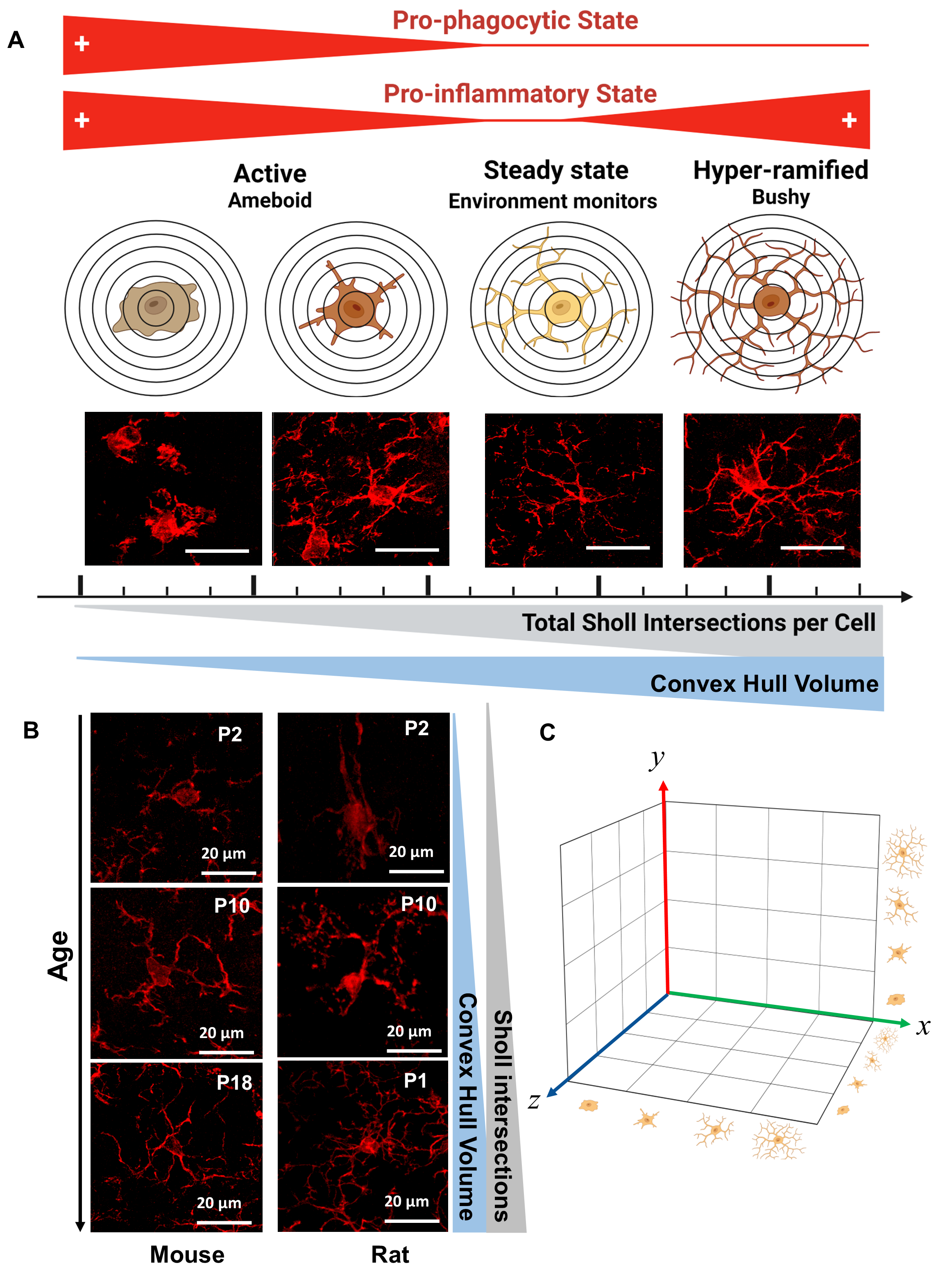
Background: The fast improvement in techniques for microstructural image capturing, processing, and analysis using sophisticated, yet reproducible and user-friendly, software has empowered researchers to quantify tridimensional whole-population cellular morphology in an unbiased manner. These technical innovations, which use immunofluorescent staining in whole-thickness brain sections have promoted novel interpretations of experimental data shifting paradigms in developmental neuroscience. Until recently microglia morphological studies have been limited to the cumbersome and still subjective process of reviewing the most common characteristics of a groups of single cells to conclude the likelihood of a “pathological” cellular milieu with the operator significantly impacting the interpretation of the images.
Objective: We hypothesized that a reproducible, blinded, and unbiased pipeline would improve our ability to detect subtle yet important differences between experimental groups.
Design/Methods:
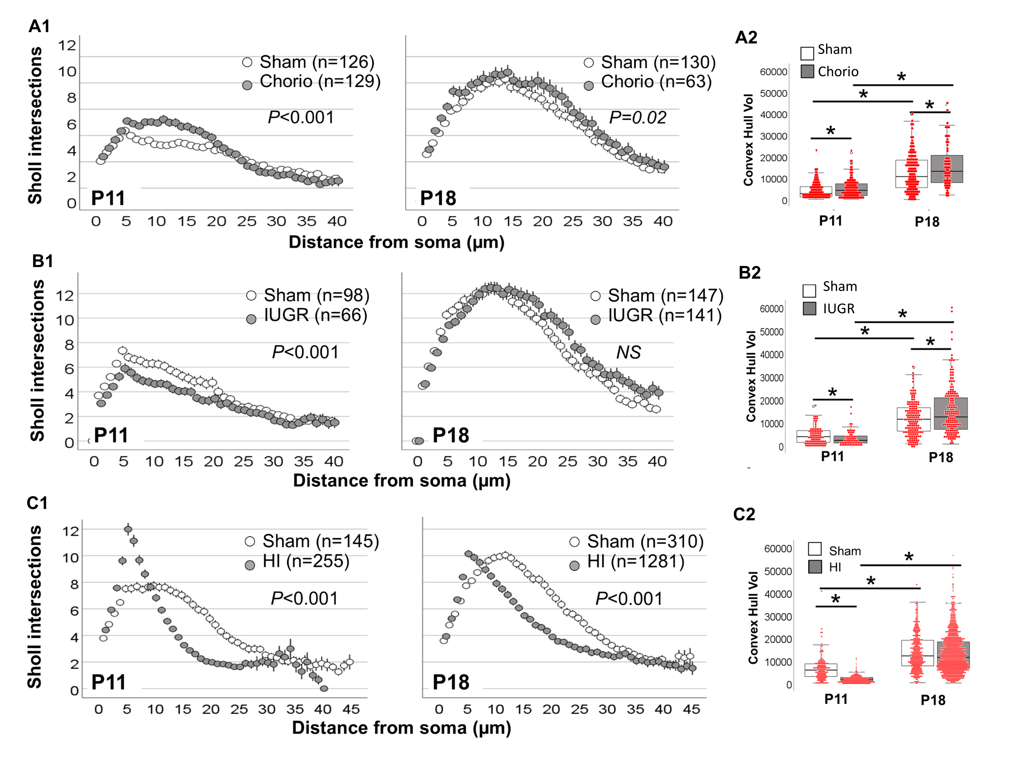
We have developed an analytical pipeline using high quality confocal microscopy and Imaris software-based techniques to address these limitations, allowing the use of highly reproducible machine-learning algorithms across all experimental repeats to quantify single-cell resolution changes within a z-stack to attenuate selection and operator biases. Thus, we evaluated the ability of our morphometric analytical pipeline to detect subtle changes in microglia populations in response to 3 separate models of developmental brain injury in rodents, these are intrauterine growth restriction (IUGR) induced in E12.5 mice, chorioamnionitis (CHORIO) induced in E16 rats, and neonatal hypoxia-ischemia (HI) induced in P10 mice.
Results: Our novel methods allowed unbiased study of whole microglia populations at a single-cell resolution, which allowed us to identify morphometric difference in Iba1+ microglia-like cells in response to all 3 models of developmental brain injury at different time points illustrating the developmental changes from P2 to P18 in the rat and P10 to P18 in the mouse.
Conclusion(s): We conclude that this unbiased analytical pipeline, which can be adjusted and applied to many other brain cells, improves the sensitivity to detect previously elusive morphological changes in microglia-like cells, which may promote specific inflammatory milieu and lead to worse outcomes and poor response to therapies.
.png)

