Neonatal Cardiac Physiology/Pathophysiology/Pulmonary Hypertension
Neonatal Cardiac Physiology/Pathophysiology/ Pulmonary Hypertension 5
115 - Abnormal amplitude integrated electroencephalography is associated with right ventricular dysfunction in neonates undergoing therapeutic hypothermia for hypoxic ischemic encephalopathy
Monday, May 1, 2023
9:30 AM - 11:30 AM ET
Poster Number: 115
Publication Number: 115.427
Publication Number: 115.427
Nadine Kibbi, University of Iowa, Iowa City, IA, United States; J Lauren Ruoss, Orlando Health Hospital, Orlando, FL, United States; Amy Stanford, University of Iowa Stead Family Children's Hospital, Iowa City, IA, United States; Rawan Al-Rawi, University of Iowa Roy J. and Lucille A. Carver College of Medicine, Iowa City, IA, United States; Stephanie S. Lee, University of Iowa Stead Family Children's Hospital, Iowa City, IA, United States; Patrick J. McNamara, University of Iowa Roy J. and Lucille A. Carver College of Medicine, Iowa City, IA, United States; Regan Giesinger, University of Iowa Stead Family Children's Hospital, Iowa City, IA, United States

Nadine Kibbi, MD (she/her/hers)
Fellow Physician
University of Iowa
Iowa City, Iowa, United States
Presenting Author(s)
Background: Right ventricular (RV) dysfunction is associated with adverse outcomes following hypoxic ischemic encephalopathy (HIE) and therapeutic hypothermia (TH); however, clinical signs may be subtle. Amplitude integrated electroencephalography (aEEG) is increasingly used to screen babies with suspected HIE for eligibility for TH. The relationship between aEEG patterns and RV dysfunction has not been studied.
Objective: To investigate RV function in neonates with normal vs abnormal aEEG patterns in the setting of HIE undergoing TH.
Design/Methods: A retrospective cohort study of term infants undergoing TH for HIE, admitted to the University of Iowa Neonatal Intensive Care Unit [07/20-06/22], was conducted. Moderate to severe HIE patients were eligible if they underwent aEEG monitoring and comprehensive targeted neonatal echocardiography (TnECHO) on the same postnatal day. Infants with congenital heart disease were excluded. RV dysfunction was defined as tricuspid annular systolic plane excursion (TASPE)≤6. A normal aEEG was considered continuous with sleep-wake cycling. aEEGs were analyzed by a single operator blind to TnECHO findings and vice versa. Univariate analysis was used.
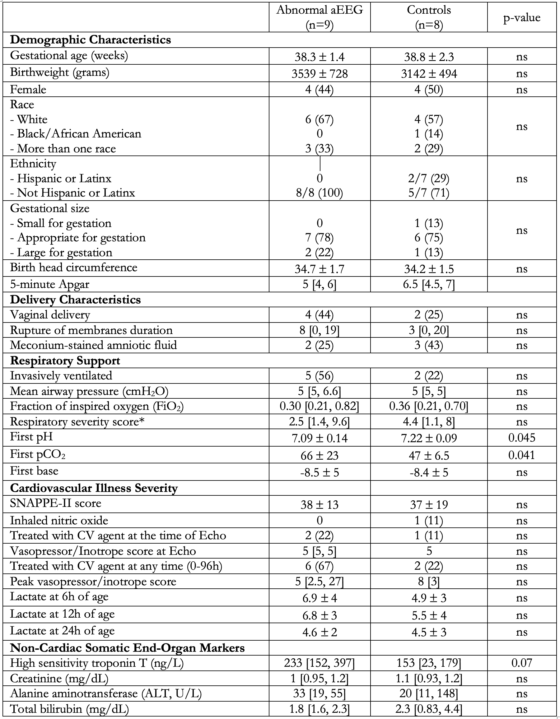
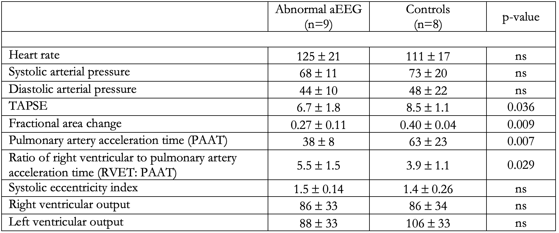
Results: A total of 17 patients were identified with either abnormal (n=9) or normal aEEG (n=8). The aEEG abnormalities were discontinuous (n=1) and absence of sleep-wake cycling (m=8). There were no differences between groups in baseline demographics, delivery characteristics, or cardiorespiratory illness severity apart from lower first pH in the abnormal aEEG group.[Table 1] There were 6 patients in the abnormal aEEG group with RV dysfunction vs 3 in the normal aEEG group (p=0.05). Markers of RV function including TAPSE (p=0.036), RV fractional area change (FAC, p=0.009), and right ventricular output (RVO, p< 0.001) were lower in the abnormal aEEG group as was low cardiac output with no difference in blood pressure or heart rate.[Table 2] Additionally, markers of pulmonary vascular tone were significantly elevated in the abnormal aEEG group.[Table 2]
Conclusion(s): Neonates undergoing TH for HIE with abnormal aEEG are more likely to have RV dysfunction. Neonates undergoing TH for HIE who demonstrate an abnormal aEEG pattern may benefit from echocardiography screening to evaluate for subclinical RV dysfunction.


