Neonatal Follow-up
NICU Follow Up and Neurodevelopment 6: The NICU Stay and Outcomes
56 - Neurodevelopmental Outcomes of Neonates with Hypoxic Ischemic Encephalopathy with Isolated White Matter Increased Diffusivity on MRI
Publication Number: 56.446
- AL
Abigail Leathe, MD (she/her/hers)
Neonatal-Perinatal Medicine Fellow
Keck School of Medicine of the University of Southern California
Los Angeles, California, United States
Presenting Author(s)
Background: Magnetic resonance imaging (MRI) is often used clinically to predict long term neurological outcome for infants with hypoxic-ischemic encephalopathy(HIE). Isolated increased diffusivity of the white matter (WM) without intrinsic T1 changes suggests injury but its implication on long-term outcome is unclear. This study aimed to compare neurodevelopmental outcomes of HIE infants with isolated increased diffusivity of WM and those with normal MRIs.
Objective:
To compare MR spectroscopy markers of injury and neurodevelopmental outcomes of HIE infants with isolated WM hyper-diffusivity versus normal brain MRIs.
Design/Methods:
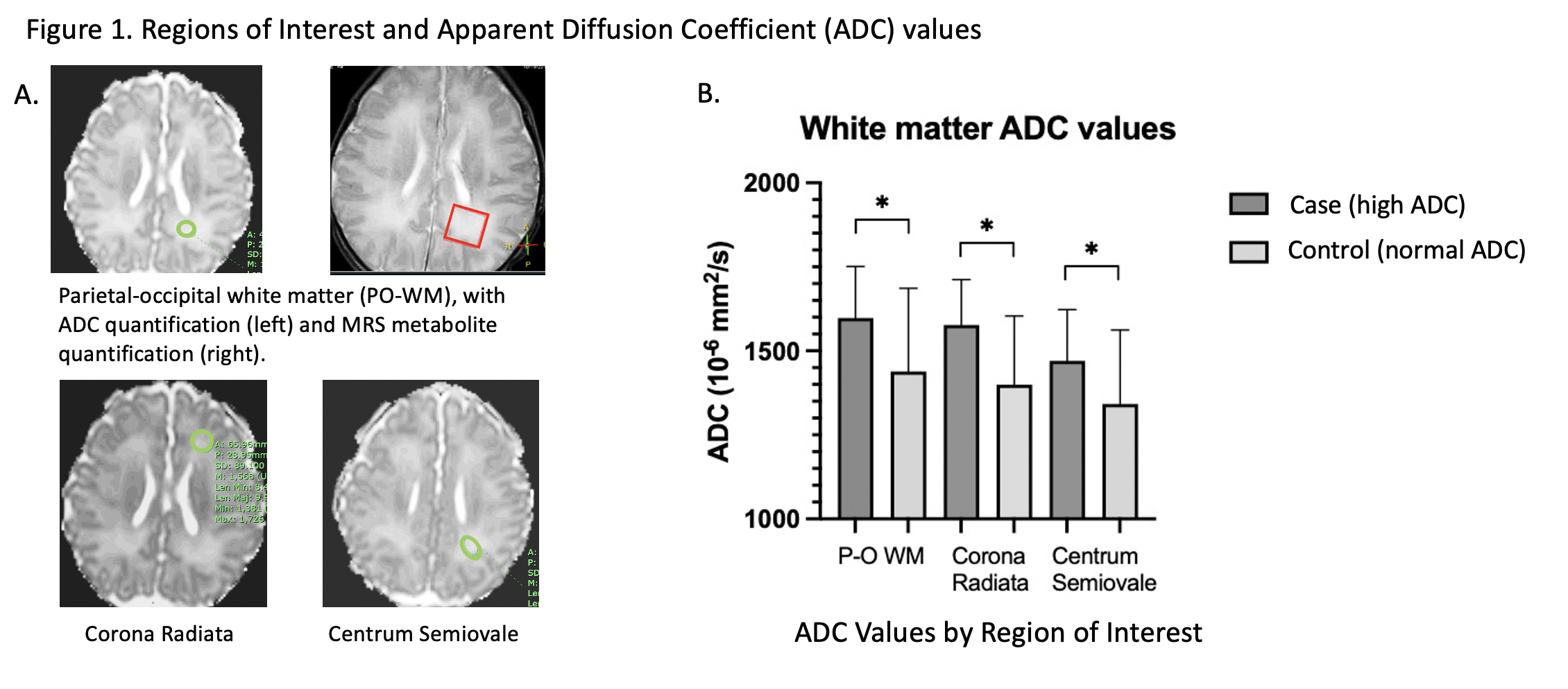
This was a retrospective review of brain MRIs of infants with suspected HIE who underwent therapeutic hypothermia between 2009 and 2019. We excluded MRIs obtained after 10 days of life to minimize effects of pseudonormalization. Case group is defined as infants with WM increased diffusivity but no correlative intrinsic T1 changes, and the control group are infants without T1,T2 and apparent diffusion coefficient (ADC) signal abnormalities on brain MRI. ADC was measured at 3 regions of interest: parietal-occipital WM, corona radiata, and centrum semiovale, Figure 1A. Cerebral metabolites related to injury (lactate, NAA, myoinositol, PUFA) were quantified using MR spectroscopy at the parietal-occipital WM. Neurodevelopmental outcomes were based upon Bayley scores at 12-24 month follow-up. Unpaired t-test, Mann-Whitney U test, and Pearson correlation were used for data analysis.
Results:
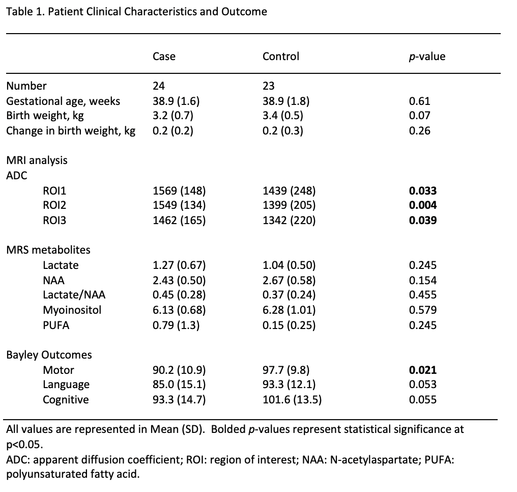
Overall, 250 brain MRIs were reviewed and 47 cases and 68 controls were identified. Twenty-four cases and 23 controls had documented Bayley scores at 12-24 months of age. Cohort characteristics are detailed in Table 1. WM ADC values were significantly higher in case group than control group in all regions, Figure 1B. For outcome, there were no difference in Bayley language (85 ±15 vs. 93±12, p >0.05) and cognitive composite scores (93±15 vs102±14, p >0.05) between case and control group, but the motor composite score was significantly lower in cases compared to controls (90±11 vs. 98±10, p=0.02), Table 1. Cerebral metabolites did not differ between case and control (all p >0.2). There was also no significant correlation between ADC and gestational age or change in birthweight at MR scan (all p >0.2).
Conclusion(s):
Isolated white matter hyperdiffusivity on brain MRI may suggest mild subacute injury as acute MRS markers of injury and T1 T2 abnormalities are absent. However, infants with isolated WM increased diffusivity may still be at risk for mild motor impairment.

