Neonatal Neurology: Clinical Research
Neonatal Neurology 5: Clinical
112 - Evaluation of neonatal brain white matter development using diffusion basis spectrum imaging
Publication Number: 112.337

Erjun Zhang (he/him/his)
PhD student
Polytechnique Montreal
Montreal, Quebec, Canada
Presenting Author(s)
Background: Diffusion tensor imaging (DTI) allows characterization of brain microstructure. However, DTI only shows averaged diffusion effects. Diffusion basis spectrum imaging (DBSI) was developed by decomposing MR signal into fiber, intracellular, extracellular and water fractions1. It has not yet been applied to preterm infant brain development.
Objective: To characterize main white matter micro-architecture in preterm by using DBSI. More specifically to: 1) Detect fiber, intracellular and extracellular fraction in preterm; 2) Assess potential differences with term controls.
Design/Methods:
Two groups of infants were scanned on a 3T MRI (2x2x2mm3, 2 b0 and 25 b (0 < b ≤ 800 s/mm2). Preterm born at 32.00 ± 1.49 weeks, scanned at 34.14 ± 1.19 weeks (G1, n=15) and at 40.18 ± 0.90 weeks (G2, n=11); 5 term control infants scanned at 39.51 ± 1.38 weeks (G3).
Data was processed by denoising, TOPUP2, registration and DBSI. Optic radiation (OR), posterior limb of internal capsule (PLIC), corpus callosum (CC), external capsule (EC) and anterior limb of internal capsule (ALIC) were manually selected on color-coded FA maps.
Statistics: comparisons (G1 vs G2, G2 vs G3) were done in ROIs using DTI and DBSI metrics (Mann-whiteney, p< 0.05).
Results:
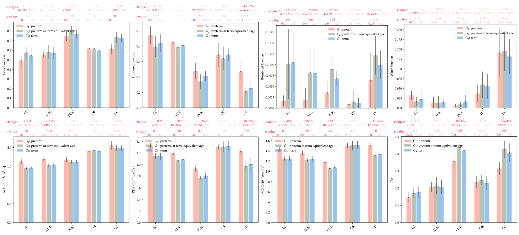
Preterm at term vs Term controls: DTI and DBSI showed no significant changes in all ROIs.
Preterm at 34 weeks vs Preterm at term equivalent age:
1. In EC, PLIC, and CC, DBSI showed drastic changes with significant fiber fraction increase (16.79%, 7.74% and 19.77%), extracellular fraction decrease (−15.83%, −28.22% and −54.76%), intracellular fraction increase (487.22%, 154.90% and 87.64%). DTI showed moderate changes: RD (−13.49%, −16.76% and −21.15%), MD (−12.51%, −10.25% and −12.81%) and FA (17.16%, 23.34% and 35.63%) (similar with previous publication3).
2. In ALIC, significant changes were found in DBSI results (324.23% intracellular fraction increase), and in DTI metrics (decrease of 9.44% for AD, 10.69% for RD, 10.17% for MD).
3. Optic radiation (OR) shows early maturation already in G1, with no major changes in both models when compared to preterm at term.
Conclusion(s): Infants, from 34 to 40 weeks, experience a major increase in intracellular fraction with moderate increase in fiber fraction. 32-week preterm infants managed to reach the same level of maturation of term control in main white matter. Optic radiation was found to reach significant maturation by 34 weeks. DBSI brings new information at the microstructural level with a granular increase.
References
1. Wang et al., Brain, 2011.
2. Andersson et al., Neuroimage 2003.
3. Kersbergen et al.,Neuroimage, 2014.
