Neonatal Follow-up
NICU Follow Up and Neurodevelopment 1: Developmental and Sensory Disorders
202 - Optic nerve head morphology in middle-aged adults born preterm with very low birth weight
Publication Number: 202.142

Maarit K. Kulmala, Medical Doctor, Ophthalmology (she/her/hers)
PhD student
FInnish Institute for Health and Welfare, Helsinki University Hospital
Espoo, Uusimaa, Finland
Presenting Author(s)
Background:
The development of neural tissue in the eye is a complex sequence of cell proliferation and apoptosis. Preterm birth affects the development of the retina and the visual tract causing retinopathy of prematurity (ROP) and visual impairment in its most severe form, but there is also evidence of more subtle structural changes. Awareness of the frequency and quality of these changes may be important for the diagnostics of degenerative eye diseases in the aging preterm population.
Objective:
The aim of this study was to characterize optic nerve head (ONH) morphology in adults born preterm with very low birth weight (VLBW; birth weight < 1500g) approaching their 40s.
Design/Methods:
From the Helsinki Study of Very Low Birth Weight Adults, 78 preterm born VLBW participants (mean gestational age 29.5 weeks, SD = 2.4) and 78 term-born controls (mean gestational age 40.0 weeks, SD = 1.1) were assessed at age 38 to 43 years. The following OHN features used in the diagnostics of optic nerve diseases were assessed by optical coherence tomography: the nerve fiber layer thickness (NFLt) in and around the ONH in six anatomical sectors, and the size of the ONH. Eyes with glaucoma or optic neuritis were excluded. None of the VLBW participants had been treated for ROP or had visual impairment. VLBW group was stratified in EP-VLBW (extremely preterm birth before gestational age 28 weeks, n=20) and non- EP-VLBW (gestational age ≥28 weeks, n= 58) subgroups. Participants (n=4) with cerebral palsy (CP) were analyzed separately.
Results:
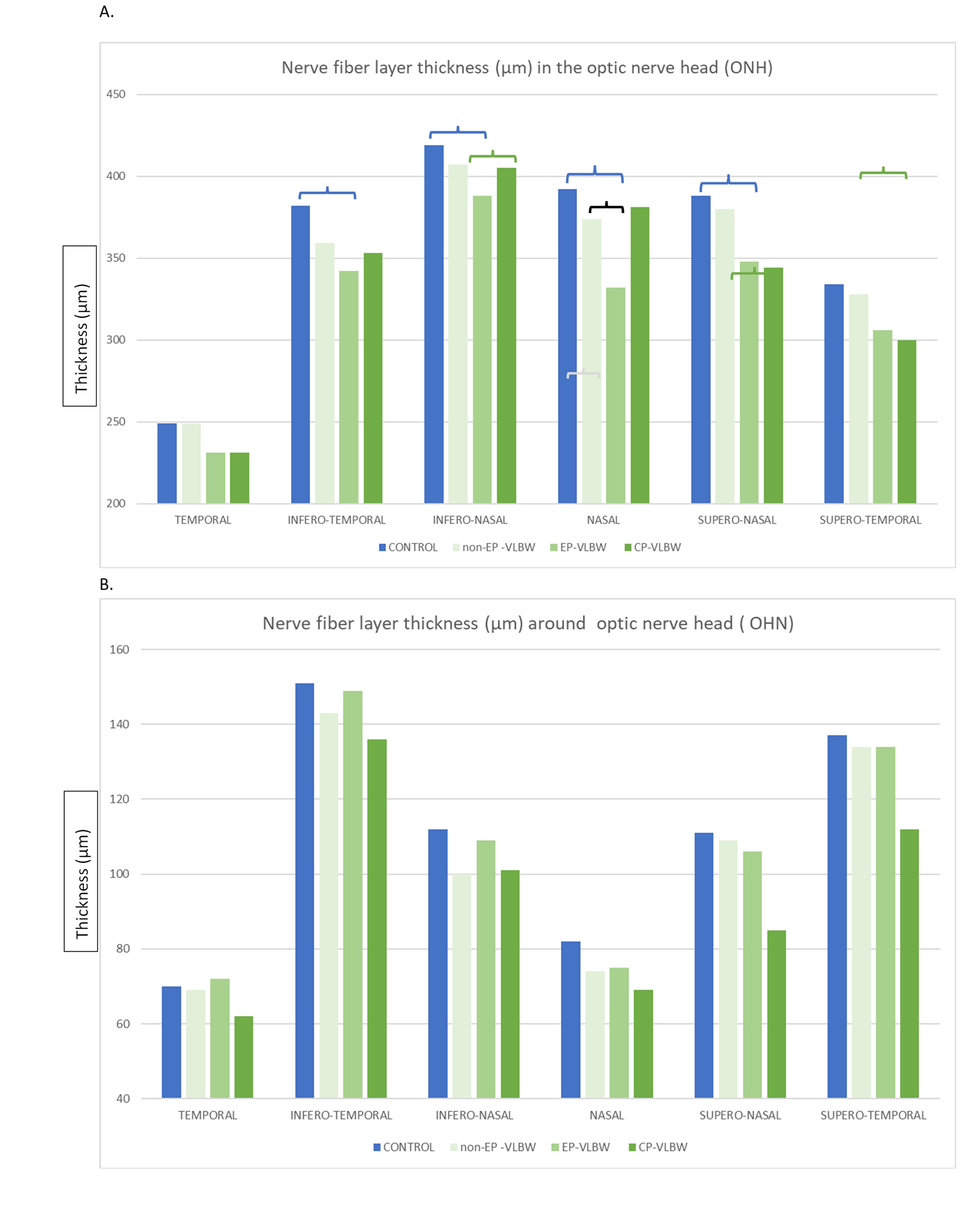
VLBW participants had less ONH neural tissue than the control eyes and with dissimilar anatomical distribution, as shown by global thinning of NFLt in and around the ONH (p< 0.05), with the most significant deviations in the nasal and inferior sectors, and by relative preservation of the temporal sector (Figure 1). EP-VLBW was associated with the thickest temporal NFLt being within or even over the high normal level. ONH size depended on the gestational age, being larger in EP than in the controls (Figure 2). CP was associated with small ONH size and severely reduced ONH neural tissue in all sectors.
Conclusion(s):
We found that adults born preterm with VLBW carried a particular ONH morphology that depended on the gestational age at birth or having CP. Alterations particularly in the inferior and nasal sectors of ONH should be appreciated while assessing any acquired ONH abnormality later in life.

.jpg)
