Neonatal Neurology: Clinical Research
Neonatal Neurology 5: Clinical
118 - Head Circumference Influences Brain Region Captured by Near-Infrared Spectroscopy in Preterm Infants
Publication Number: 118.337

Sarah E. Kolnik, MB MBA (she/her/hers)
Assitant Professor of Pediatrics
University of Washington - Seattle Children's Hospital
Seattle, Washington, United States
Presenting Author(s)
Background:
Cerebral Near-Infrared Spectroscopy (cNIRS) is a non-invasive clinical tool used to measure regional cerebral tissue oxygenation (rScO2), created and validated in adult and pediatric populations. Given their vulnerability for neurologic injury, preterm neonates are attractive candidates for cNIRS; however, normative data and reference to the brain regions measured by cNIRS have not yet been established for this population.
Objective:
The aim of this study was to analyze continuous cNIRS readings within the first 6-72h after birth in preterm neonates to determine if cNIRs readings are altered by head circumference (HC) or brain regions captured.
Design/Methods:
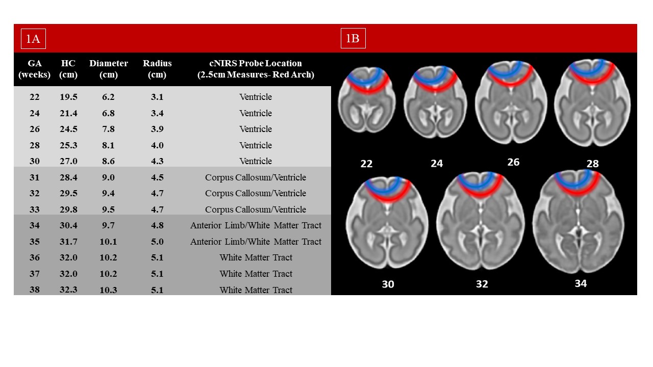
This is an observational single center cohort study of infants born ≤ 1250 g and/or ≤ 30 weeks’ gestation. Infants were monitored prospectively with a continuous infant-neonatal cNIRS (INVOSTM) probe that provides rScO2 at a depth of ~2.5 cm. Brain regions captured at this depth were compared to a published fetal T2 MRI brain atlas. Demographic and clinical data collection occurred retrospectively. Infants with congenital anomalies, head circumference > 30 cm, intracranial hemorrhage, or death prior to 72h were excluded. Descriptive statistics were used to describe the demographic variables of the included infants using either median and interquartile range (IQR) or count and percentage. NIRS curves were generated using generalized additive model (GAM) fitting with start time of 6h after birth. Trajectories of cNIRS rSO2 were plotted using GAM based on HC and gestational age (GA). For GA plots, infants with a HC Z-score more than +/- 2 were excluded.
Results:
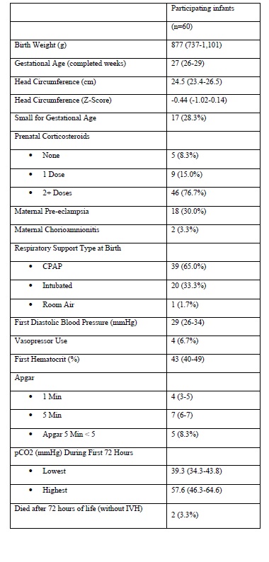
During the study, 98 infants were monitored with cNIRS. Sixty infants (61.2%) were included in this analysis. Baseline characteristics of the study population are depicted in Table 1. In infants with HC < 30 cm, a portion of the cNIRS readings reflects ventricular CSF (Figure 1). For infants with HC < 27cm, the majority of the cNIRS reading comes from ventricular CSF and deep brain structures (Figure 1). cNIRS readings were associated with both GA and HC; the most immature infants and those with the smallest HCs had the lowest measured rScO2 trajectories (Figure 2A, 2B). After stratifying for GA, a smaller HC was still associated with lower rScO2 (Figure 2C, 2D).
Conclusion(s):
This study highlights the importance of rigorously re-validating technologies before extrapolating them to different populations. Clinicians should be aware that in preterm infants with small HCs, rScO2 displayed may not reflect oxygenation in cerebral tissue as the signal is largely derived from CSF in the ventricle. 

.jpg)
