Neonatal Quality Improvement
Neonatal Quality Improvement 3
729 - Transhepatic and Subcostal Ultrasound Imaging for Catheter Location of Subcutaneously Tunneled Mid-Thigh Femoral Vein Catheters in Neonates
Publication Number: 729.342

Matthew D. Ostroff, MSN, APN (he/him/his)
Vascular Access Coordinator / Lead Nurse Practitioner
St. Joseph's Children's Hospital
Paterson, New Jersey, United States
Presenting Author(s)
Background:
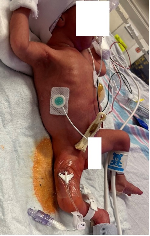
Our 2020 publication in the Journal of Vascular Access showed that subcutaneously tunneled femorally inserted central catheter (ST-FICC) (Fig 1) placement is an important procedure to provide parenteral nutrition and medications to critically ill neonates while preserving their upper extremities vasculature. [1] We currently confirm FICC position using the standard radiography.
Objective:
To improve patient care and decrease radiation exposure to the neonates, we recently adapted the use of point-of-care-ultrasound (POCUS) to confirm neonatal FICC placement. We compared the accuracy of POCUS to standard radiography for verification of vessel and catheter terminal tip location.
Design/Methods:
A quality improvement (QI) initiative was performed in Dec 2022 on twenty neonates (Gestational Age: 23-38 weeks; Birth Weight 510-3130g) requiring placement of a 1.4 or 1.9 French ST-FICC in a level 3, 30-bed NICU. The procedure was performed by a vascular advanced practice nurse under ultrasound guidance. Following placement, transhepatic ultrasound view (Fig 2) using a 12-6 linear probe at 6cm depth was performed to identify the aorta, inferior vena cava (IVC), and hepatic veins. Catheter placement above the level of the hepatic vein within the IVC confirmed venous placement. A subcostal ultrasound view at 3.1 cm depth was then performed identifying the right atrium (RA) to visualize “crystals” (Fig 3) following a saline flush indicating the tip position in the upper one third of the IVC. Two views of the chest and abdomen radiographs were then performed to verify catheter location and tip positioning.
Results:
POCUS guided venous vessel confirmation was 100% compared to radiography. In one case, POCUS was superior to x-ray in defining that FICC was in IVC due to neonate’s abdominal distention. POCUS catheter tip location in the IVC was 94% accurate compared to radiograph. In one case, the catheter terminated within the RA and the saline flush was not visualized with ultrasound. POCUS allowed for real time verification of FICC placement.
Conclusion(s):
POCUS is an effective and efficient way to localize central venous tip line position for ST-FICC in neonates. It is a safe, portable, ionizing radiation free alternative to standard radiography. A prospective, investigational powered study is currently ongoing to validate this QI project.
1. Ostroff, M., Zauk, A., (2021). A retrospective analysis of the clinical effectiveness of subcutaneously tunneled femoral vein cannulations at the bedside. The Journal of Vascular Access, 22(6), 926-934.
B02BD055-42D0-42D2-8579-B7DB9440D87A.jpeg

80D770D1-C05A-47E2-8060-D80E99E00534.jpeg
