Neonatal Follow-up
NICU Follow Up and Neurodevelopment 1: Developmental and Sensory Disorders
201 - Association Between Preterm Infant Retinal Microanatomy at 34-38 Weeks Postmenstrual Age and Neurodevelopment at 2 Years of Age
Publication Number: 201.142

Kathryn E. Gustafson, PhD (she/her/hers)
Professor
Duke University School of Medicine
Durham, North Carolina, United States
Presenting Author(s)
Background: Preterm infants are at increased risk for adverse neurodevelopmental outcomes with worse outcomes in infants with unfavorable visual status. It is important to clarify the relationship between retinal microanatomy, visual pathway development, and later neurodevelopmental functioning.
Objective: To evaluate the association between optical coherence tomography (OCT)-based retinal microanatomical findings in the nursery and 2-year neurodevelopmental measures in preterm infants.
Design/Methods:
As part of BabySTEPS (STudy of Eye imaging in Preterm infantS), infants at risk for retinopathy of prematurity (ROP) had bedside imaging in the nursery with an investigational OCT system. Thickness of retinal nerve fiber layer (RNFL) near the optic nerve and retinal layers at the foveal center were measured from OCT images at 34-38 weeks postmenstrual age (PMA). Neurodevelopmental assessments at 2-years corrected age included cognitive, language, and motor scales of the Bayley Scales of Infant and Toddler Development – 3rd ed. (BSID) and the parent rated Child Behavior Checklist (CBCL) and Modified Checklist for Autism in Toddlers-Revised (MCHAT-R). Multivariable linear regression models were used to evaluate associations between the measures of retinal microanatomy and of 2-year neurodevelopment, with adjustment for birth weight, gestational age, PMA of imaging, and mother’s highest education level.
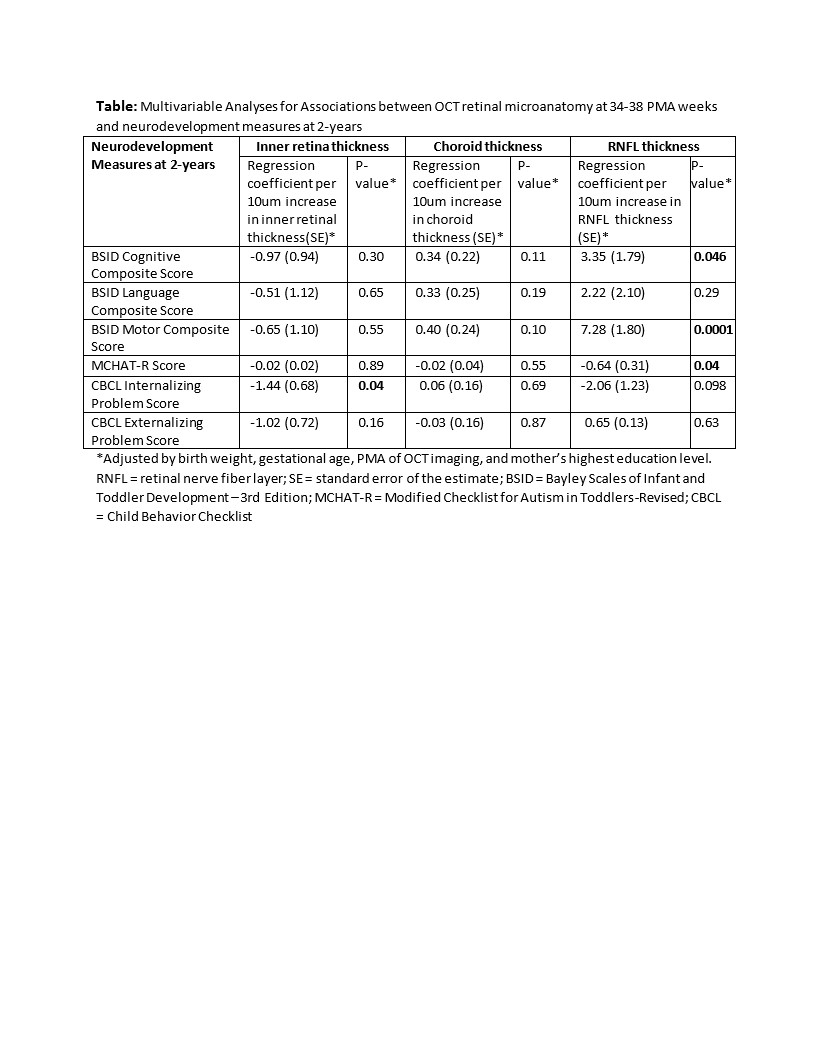
Results: Of 98 children with ≥1 OCT imaging session in 34-38 weeks PMA, 72 returned for the 2-year assessment. In multivariable analysis (see Table), RNFL thickness was associated with BSID Cognitive (p=0.046) and Motor (p=0.0001) composite scores, and MCHAT-R score (p=0.04). Greater inner retina thickness was associated with lower CBCL Internalizing problems score (p=0.04). In univariable analysis, greater choroid thickness was associated with higher BSID Cognitive (p=0.0004), Language (p=0.004), and Motor (p=0.0004) composite scores, and lower MCHAT score (p=0.04); but these did not remain significant with multivariable analysis.
Conclusion(s):
In preterm infants, greater RNFL thickness at 34-38 weeks PMA was associated with higher cognitive and motor development scores and lower autism risk scores at age 2. Thinner RNFL in preterm infants has previously been associated with poorer visual acuity at 9 months. The current findings point to the potential for RNFL (comprised of axons which extend to the lateral geniculate nucleus) as a marker either of parallel neurodevelopment, or of visual function and its impact on neurodevelopment in preterm infants.
