Neonatal Neurology: Pre-Clinical Research
Neonatal Neurology 9: Preclinical 3
40 - Assessment of cerebral perfusion and metabolism following neonatal seizures in a preclinical model
Publication Number: 40.435
- SM
Subhabrata Mitra, PhD (he/him/his)
Associate Professor and Honorary Consultant Neonatologist
University College London
London, England, United Kingdom
Presenting Author(s)
Background:
Seizures induce significant changes in cerebral metabolism and perfusion. MR imaging and proton MR spectroscopy (1H MRS) are standard MR tools to assess neonatal seizures. Phosphorus (31P) MRS and arterial spin labelling (ASL) MRI are other techniques for the assessment of cerebral metabolism and perfusion respectively.
Objective:
To assess the effect of seizures on brain perfusion and metabolism using pre and post ictally acquired MR imaging, 1H MRS, 31P MRS and ASL MRI, in a preclinical model of a healthy newborn brain.
Design/Methods:
Ten healthy male newborn piglets had seizure induction with bicuculine 4mg/kg IV over two time periods 3h apart. Piglets were sedated and anaesthetised throughout the experiment and underwent aEEG/EEG and systemic physiological monitoring. All imaging data were acquired in a 3T Philips Achieva scanner using a Lammers Medical MR Conditional transport incubator, Lammers 8ch custom-made proton coil and Philips phosphorous flex coil. Each animal had a scan at the start of the study (pre-ictal) and a repeat scan 5h p seizure (post-ictal).
ASL was performed using pseudo-continuous labelling (pCASL) with 1.8s labelling duration and 2s post-labelling delay and a 3D GRASE readout (TE/TR=13 ms/4187 ms, voxel size 2.5*2.5*5 (mm), 26 slices). Cerebral blood flow [ml/100g/min] maps were computed and mean CBF[ml/100g/min] within regions were quantified using an automated atlas segmentation approach. Chemical shift imaging (CSI) was acquired with an 8 × 8 matrix, a voxel size of 7.5 × 7.5 mm2 and slab thickness of 10 mm (TE/TR= repetition time was 288 ms / 2 s. CSI data was processed using TARQUIN yielding signal amplitudes. 31P MRS was acquired with Image-Selected In vivo Spectroscopy (ISIS) voxel size of 40x55x70 mm (adapted to include the whole brain) (FID, TR=10000ms). ISIS data was processed using JMRUI.
Results:
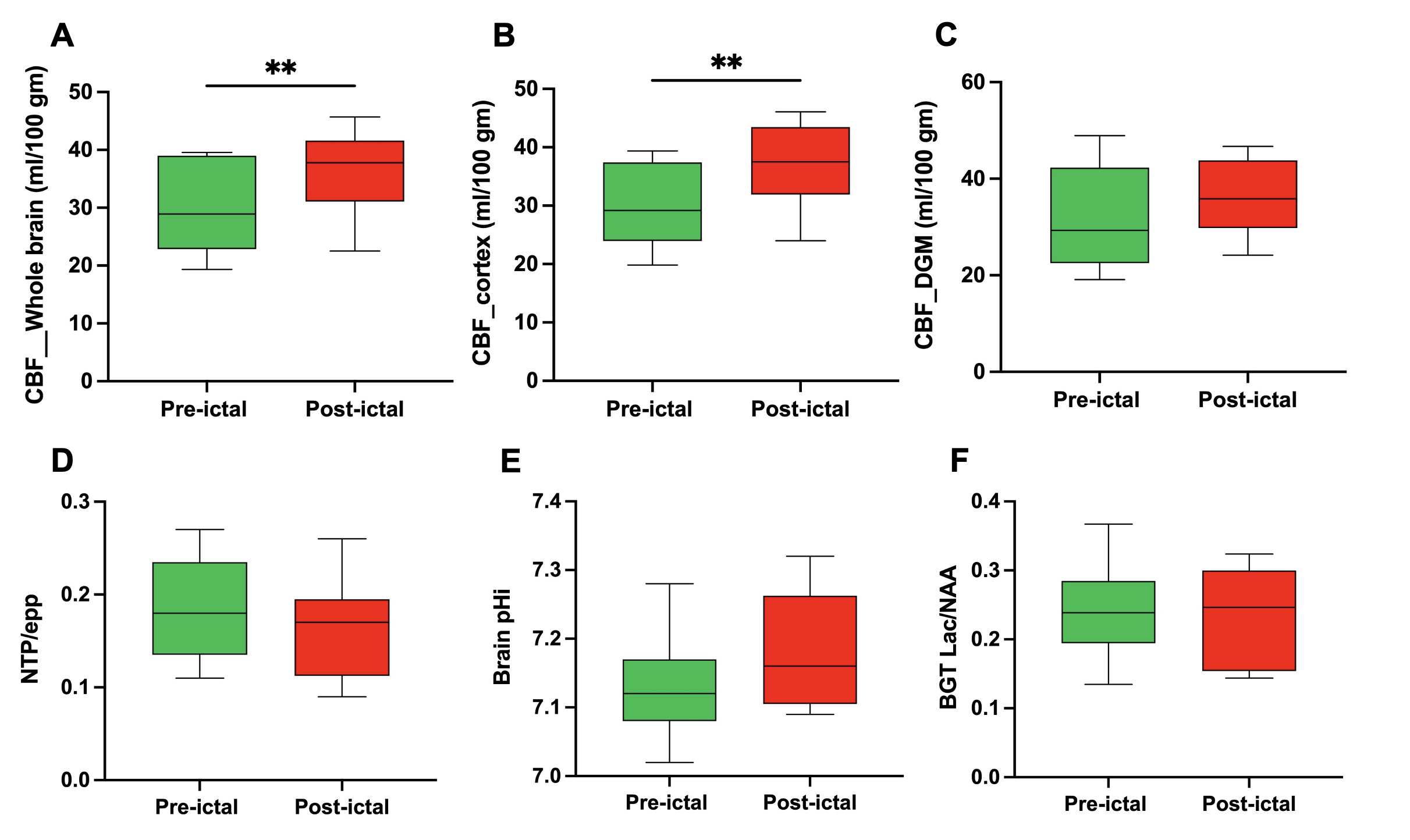
Generalised tonic clonic seizures with electrographic changes noted immediately after bicuculine administration. Significant differences noted between the pre-ictal and post-ictal period in the whole brain (P=0.007) and cortical (P=0.004) blood flow (CBF) (figure 1A, 1B). In the post-ictal period, there was a trend to an increase in Intracellular pH (pHi) as a result of postepileptic intracellular alkalosis (fig 1E), (7.13±0.07 vs 7.18±0.08) and reduction in brain energy metabolism high energy phosphate state (NTP/epp) (figure 1D). Lac/NAA on 1H MRS did not differ between the two scans.
Conclusion(s):
Increased whole brain and cortical perfusion measured on ASL-MRI persisted following seizures 5h after the seizure induction in a preclinical seizure model.

