Neonatal Neurology: Pre-Clinical Research
Neonatal Neurology 9: Preclinical 3
31 - Delta Like 4 is Required for Perinatal Brain Vascularization and Repressed by Postnatal Hyperoxia in Mouse Brain Endothelial Cells
Publication Number: 31.435

Xingrao Ke, MD (she/her/hers)
Research Scientist
Children's Mercy Hospital
Kansas City, Missouri, United States
Presenting Author(s)
Background: The cerebral vasculature expands rapidly to support neurogenesis and brain development during the perinatal period. The molecular mechanisms regulating brain vascularization during perinatal development are not fully understood. Further, exposure to high concentrations of oxygen in premature infants impairs lung and retinal vascular development, and likely cerebral vascular development. In this study we investigated whether delta like 4 (Dll4), a Notch ligand that is important for brain vascularization, is affected by postnatal hyperoxia exposure in the endothelial cells (ECs) of developing brain. We hypothesized that postnatal hyperoxia exposure decreases Dll4 expression in mouse brain ECs.
Objective: Postnatal hyperoxia exposure decreases Dll4 expression in mouse brain ECs.
Design/Methods:
We compared vascular phenotype and Dll4/Notch signaling gene levels in brain ECs between wild type (Dll4+/+) mice and LacZ reporter (Dll4+/LacZ) mice that lack one allele of Dll4. We compared gene expression in immortalized human brain microvascular ECs (HBMECs) treated with Dll4-shRNA to scramble RNA controls. We performed in vitro angiogenesis assay, trans-endothelial electrical resistance (TEER) measurement and in vitro vascular permeability assay. We compared Dll4 expression in the ECs on postnatal day (P) 15 mouse brain exposed to 85% hyperoxia or room air between P1-14.
Results:
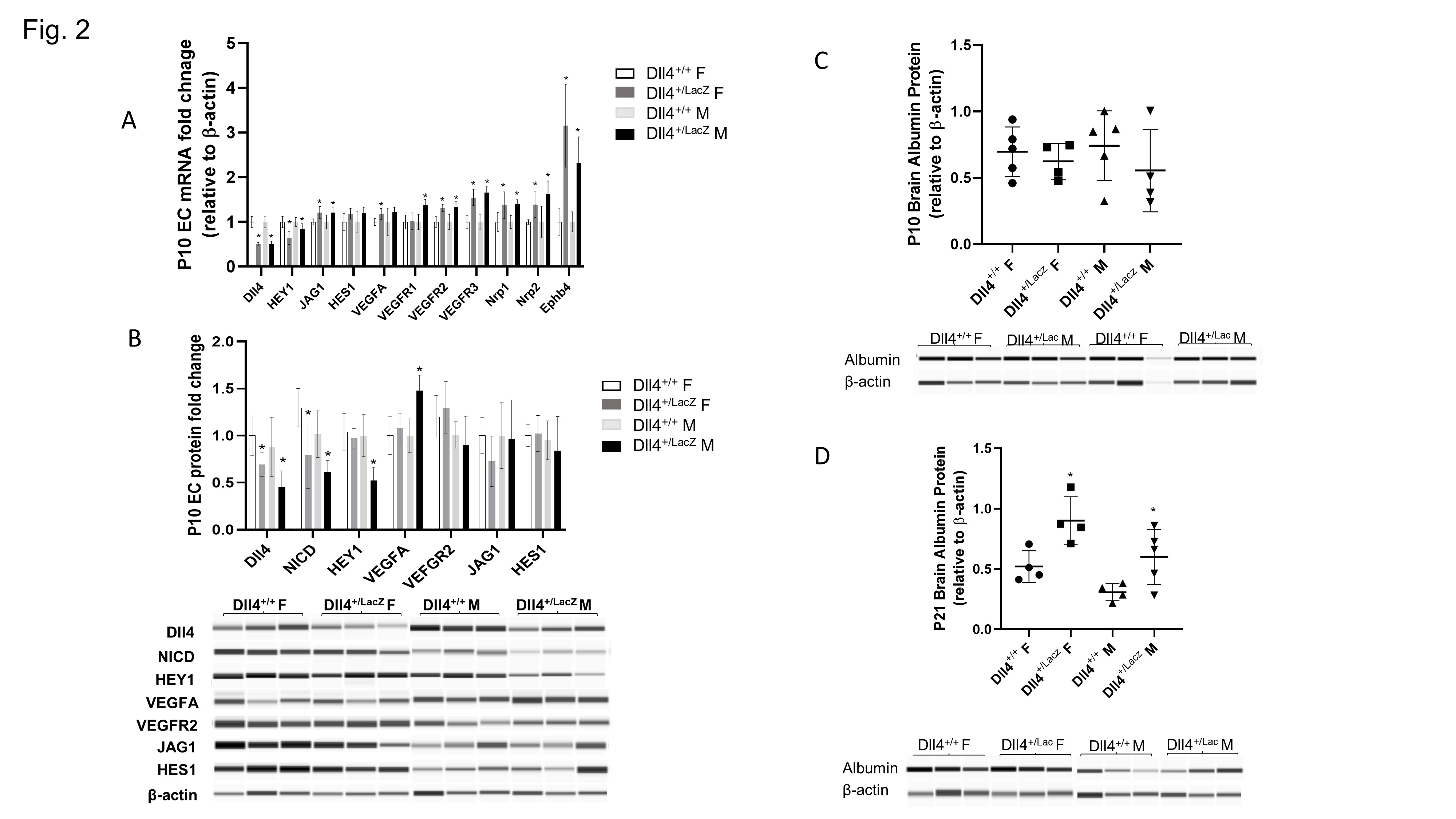
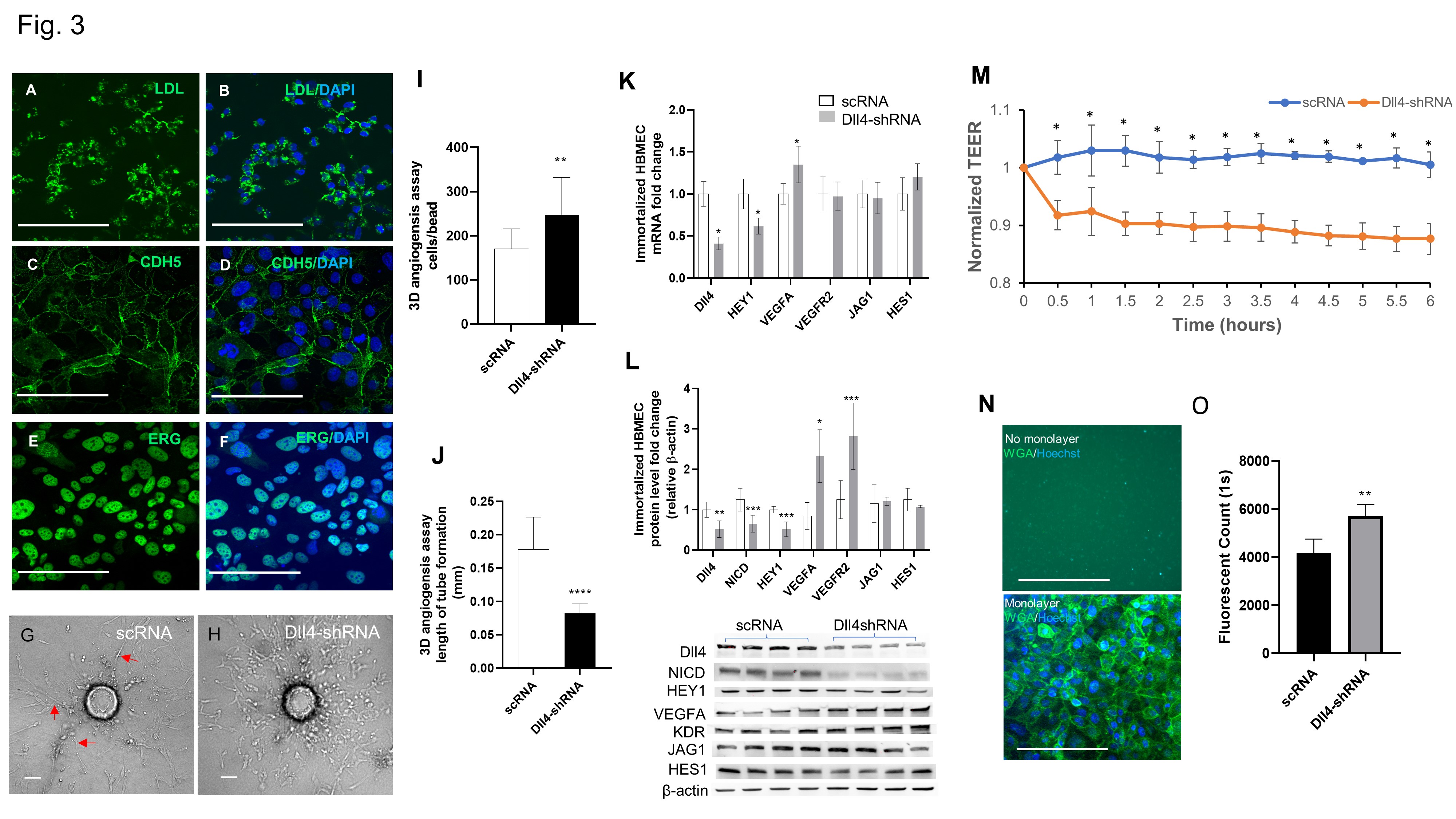
Dll4 was prominently expressed in arterial ECs in mouse brains at P7 and P21, and in human brains at 39-week gestation and 4-year. Dll4 haploinsufficiency resulted in dysmorphic vasculature evidenced by decreasing capillary diameter in developing and adult brains in a sex-specific manner (Fig.1). Dll4 insufficiency decreased Dll4-NICD-HEY1 signaling in Dll4+/LacZ brain ECs in vivo and in Dll4-shRNA treated HBMECs in vitro (Fig. 2&3). Furthermore, Dll4 insufficiency induced deviant angiogenesis in vitro, increased the permeability of HBMEC and impaired BBB integrity in Dll4+/LacZ mice (Fig.3). Postnatal hyperoxia exposure decreased Dll4 expression in the ECs of developing mouse brain.
Conclusion(s):
We conclude that Dll4 regulates cerebral vascular development through Dll4-NICD-HEY1 signaling and postnatal hyperoxia exposure downregulates Dll4 expression in the ECs of developing mouse brain. We speculate that decreased Dll4 expression in brain ECs may play a role in poor neurodevelopmental outcome seen in the individuals exposed to postnatal hyperoxia..jpg)


